
Thoracic Surgery Newsletter — February 2025
February 15, 2025Thoracic Surgery Newsletter — August 2025
August 15, 2025Thoracic Surgery Newsletter — May 2025

Letter from the Chief
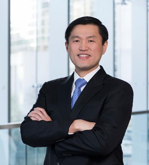
Min P. Kim, MD, FACS It is with great pride and gratitude that I reflect on the remarkable journey of robotic thoracic surgery at Houston Methodist Hospital. What began as a vision to elevate surgical care took its first step in 2011 when I performed my very first robotic case. Today, I am humbled to share that I recently reached a personal milestone-completing my 2000th robotic thoracic surgical procedure. Over the years, robotic surgery has transformed from an innovative option to a core component of our thoracic surgical program. We have advanced from routine cases to performing some of the most complex procedures with this technology, including robotic lobectomies for lung The benefits of this platform have been extraordinary. For our patients, it means less postoperative pain, shorter hospital stays, and faster recoveries. For surgeons, it means enhanced visualization, superior dexterity, and greater precision-all of which translate into safer operations with fewer complications.
Bringing robotic thoracic surgery to our community and transforming the way we care for patients could not have been possible without the unwavering support of many. I am deeply thankful for our hospital leadership and department chair for believing in this vision. I am especially grateful to our dedicated Walter Tower nurses, operating room technicians, anesthesiologists, and my surgical partners-each of whom has played a vital role in building and advancing this program. As we look to the future, I remain excited about the continued evolution of robotic surgery and our division’s role in pushing the boundaries of what is possible in thoracic care. Min P. Kim, MD, FACS
Incorporating Digital Chest Tube Technology to Enhance Postoperative Care
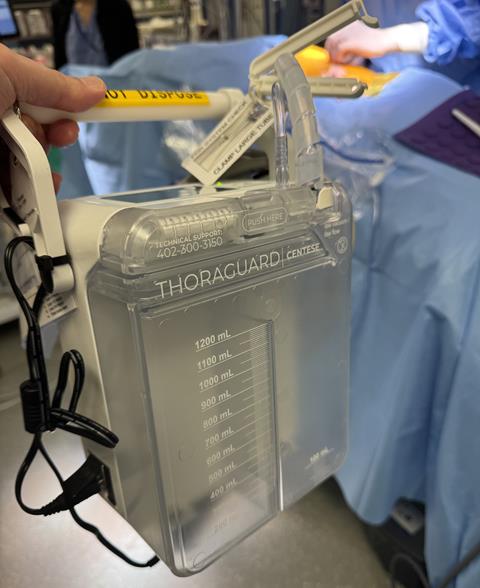
The Division of Thoracic Surgery continues to lead in advancing patient care through the thoughtful integration of new technologies. One of the latest additions to our clinical practice is the Centese digital chest tube system, which has significantly transformed the management of chest tubes for patients undergoing lung surgery. Following thoracic procedures, chest tubes are routinely placed to drain fluid and monitor for air leaks. One of the main reasons for keeping a chest tube in place after lung resection is concern for a persistent air leak from the lung parenchyma. If a chest tube is removed while a leak remains undetected, the patient is at risk of
Historically, assessing the presence and severity of an air leak has been challenging and often relied on subjective interpretation using analog systems. The introduction of the Centese digital chest tube has fundamentally changed this process by providing real-time, accurate assessment of air leak status. This enhanced precision allows for more confident and timely decision-making regarding chest tube removal. As a result, we have been able to safely remove chest tubes much earlier – in select cases, even in the recovery room immediately after surgery. This has significant benefits for our patients. Chest tubes are often one of the most painful components of We would like to extend our gratitude to Anne Louise Stansa, Rose McCullough and Tony DeDominico whose efforts were instrumental in bringing this advanced technology to Houston Methodist Hospital and integrating it seamlessly into our thoracic surgery pathway. By embracing innovations like the Centese digital chest tube, we continue to push forward in our mission to deliver the highest quality and most compassionate care for patients with thoracic surgical disease.
Division of Thoracic Surgery Spotlight
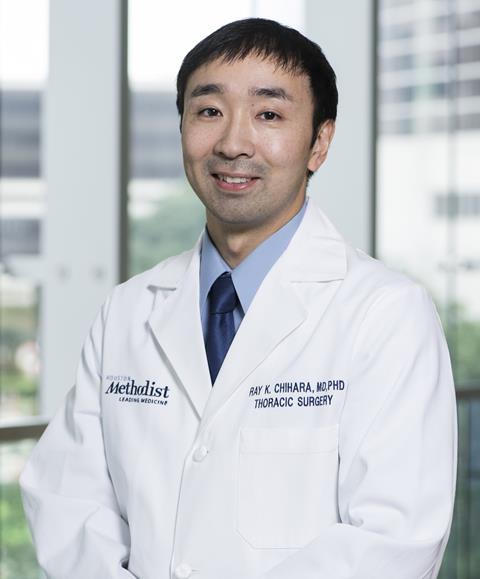
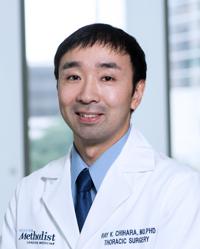
Ray K. Chihara, MD, PhD Dr. Ray Chihara: Advancing Lung Transplant Care at Houston Methodist The Division of Thoracic Surgery at Houston Methodist Hospital is proud to highlight the outstanding contributions of Dr. Ray Chihara, who has been leading the lung transplant program with dedication, innovation, and excellence in the Division of Thoracic Surgery. Bringing advanced expertise from his fellowship training at Emory University, Dr. Chihara has become an integral part of the lung transplant team. He currently performs approximately half of all lung transplants at Houston Methodist, often collaborating closely with Dr. Warren Naselsky to deliver exceptional surgical care.
Recognizing his leadership and vision, Dr. Chihara has become the driving force behind lung transplantation within the Division of Thoracic Surgery. His efforts have strengthened the program and established it as a vital part of our clinical mission. Beyond the operating room, Dr. Chihara has been instrumental in enhancing the overall care experience for lung transplant patients. Recognizing the challenges of postoperative pain in this population, he has introduced the use of cryoablation to improve pain control following transplant surgery. This approach has made a meaningful difference in patient comfort and recovery. In addition, Dr. Chihara has placed a strong focus on the long-term success of lung transplant recipients. One of his priorities has been addressing reflux and gastroparesis, known contributors to chronic lung transplant rejection. He has led efforts to prevent reflux-related complications by performing fundoplication procedures in select patients to reduce reflux and lower the risk of chronic rejection. Through careful management and preventative strategies, he is working to extend the life and function of transplanted lungs. Thanks to Dr. Chihara’s leadership and clinical expertise, the lung transplant program continues to grow and evolve, offering patients comprehensive and We are proud to recognize Dr. Chihara’s contributions and leadership and look forward to the continued advancement of lung transplant care under his guidance.
C-arm 3D Imaging Improves Diagnostic Yield of Robotic Shape-Sensing Transbronchial Biopsy
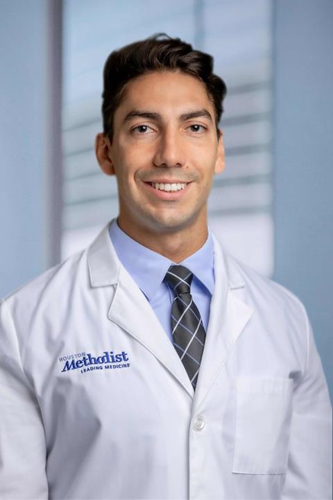
Tomasso Cambiaghi, MD We are pleased to announce that a manuscript from our division has been accepted for publication, marking another important contribution to the growing field of minimally invasive thoracic diagnostics. The study, led by Dr. Cambiaghi, Thoracic Surgery Fellow, and titled “C-arm 3D Imaging Improves Diagnostic Yield of Robotic Shape-Sensing Transbronchial Biopsy,” explores the role of advanced imaging in improving the diagnostic accuracy of robotic navigational bronchoscopy for lung nodules. As lung cancer screening becomes more widespread, detecting and accurately diagnosing small, suspicious nodules has become critically important. In our program, shape-sensing robotic navigational bronchoscopy is performed using the ION robotic platform, which offers precise navigation to peripheral nodules. This study investigated how integrating C-arm 3D imaging with the ION platform could further enhance this technique and improve diagnostic confidence.
In this single-center retrospective study, 95 patients underwent robotic bronchoscopy combined with intraoperative C-arm 3D imaging. The nodules biopsied were typically small (median size 1.7 cm) and frequently located in the upper lobes. Impressively, the procedure demonstrated minimal complications, with only one patient experiencing pneumothorax, which was managed conservatively without chest tube placement. The real strength of the approach, however, was in its diagnostic performance. The study achieved a 94% diagnostic accuracy at 12 months, with malignancy confirmed in over half of cases and benign diagnoses supported by long-term follow-up in others. This important research could not have been possible without the hard work and dedication of our team, especially Dr. Cambiaghi, who led the project with scientific rigor and clinical insight. Congratulations to all involved in this achievement and for continuing to advance the field of thoracic surgery through innovation and scholarship.
Showcasing Innovation at The American Association for Thoracic Surgery Annual Meeting
At the recent AATS Annual Meeting in Seattle, Dr. Mahnoor Zia on behalf of the Division of Thoracic Surgery presented an important advancement in robotic esophageal surgery. The presentation, titled “Enhancing Conduit Straightness: Novel Pleural Closure Technique in Robotic Ivor Lewis Esophagectomy,” focused on a technical innovation that addresses a key challenge in esophageal reconstruction. In robotic Ivor Lewis esophagectomy, ensuring the gastric conduit remains straight and unobstructed as it courses through the posterior mediastinum is critical for optimal postoperative outcomes. Kinking or displacement of the conduit can contribute to delayed 
Mahnoor Zia, MD
The study highlights a simple yet highly effective pleural closure technique, performed after conduit placement, which helps maintain the straight position of the conduit. By closing the pleural space around the conduit in a controlled manner, surgeons can prevent lateral shift or sagging of the conduit into the pleural cavity. The video presentation at AATS provided a step-by-step demonstration of this approach, illustrating the technical ease and reproducibility of the maneuver using the robotic platform. Early outcomes are promising. Patients who underwent esophagectomy using this pleural closure technique demonstrated: Fewer conduit-related complications, This innovative technique reflects the division’s ongoing commitment to refining surgical approaches to improve outcomes and enhance patient recovery. As robotic platforms continue to evolve, integrating thoughtful techniques such as pleural closure will be key to optimizing complex thoracic procedures. The Division of Thoracic Surgery remains at the forefront of clinical excellence and surgical innovation, and was honored to share this important work with peers from around the
Division of Thoracic Surgery Educational Highlight: Emergency Thoracotomy Course
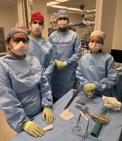
Emergency Thoracotomy Simulation Course Prepares Fellows for Critical Situations At the Division of Thoracic Surgery, advancing education is core to our mission – and that includes preparing our trainees for the unexpected. This quarter, our thoracic surgery fellows participated in a unique and essential training event: the Emergency Thoracotomy Simulation Course, held at the world-class MITIE facility at Houston Methodist Hospital (HMH).
The course, led by Dr. Min Kim, was designed to address an important educational gap. As our team continues to push the boundaries of complex robotic surgery, including robotic lobectomy for lung cancer and other advanced procedures, many of today’s trainees may not frequently encounter scenarios that demand rapid conversion to open surgery. One of the most critical and high-risk situations in thoracic surgery – a major vascular injury – requires decisive action and the ability to perform an emergency thoracotomy. During this immersive course, current cardiothoracic surgery fellows Drs. Anuj Shah and Tommaso Cambiaghi performed simulated robotic lobectomies The simulation was made as realistic as possible thanks to the outstanding support from Patty Phung, RN, BSN, CNOR and Ashley Pozo, CST, members of the Walter Tower Operating Room staff, who meticulously recreated an authentic operating room environment. Their expertise helped ensure that the fellows were able to engage fully in a setting that closely mimicked real-life operative conditions. We are By proactively preparing our fellows for rare but critical events, we ensure that they graduate from our program not only as technically skilled robotic surgeons, but also as well-rounded, confident, and safe thoracic surgeons capable of managing any intraoperative challenge.
Ray K. Chihara, MD, PhD 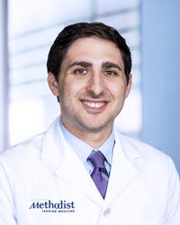
Warren C. Naselsky, MD, MS 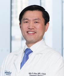
Min P. Kim, MD

|
